
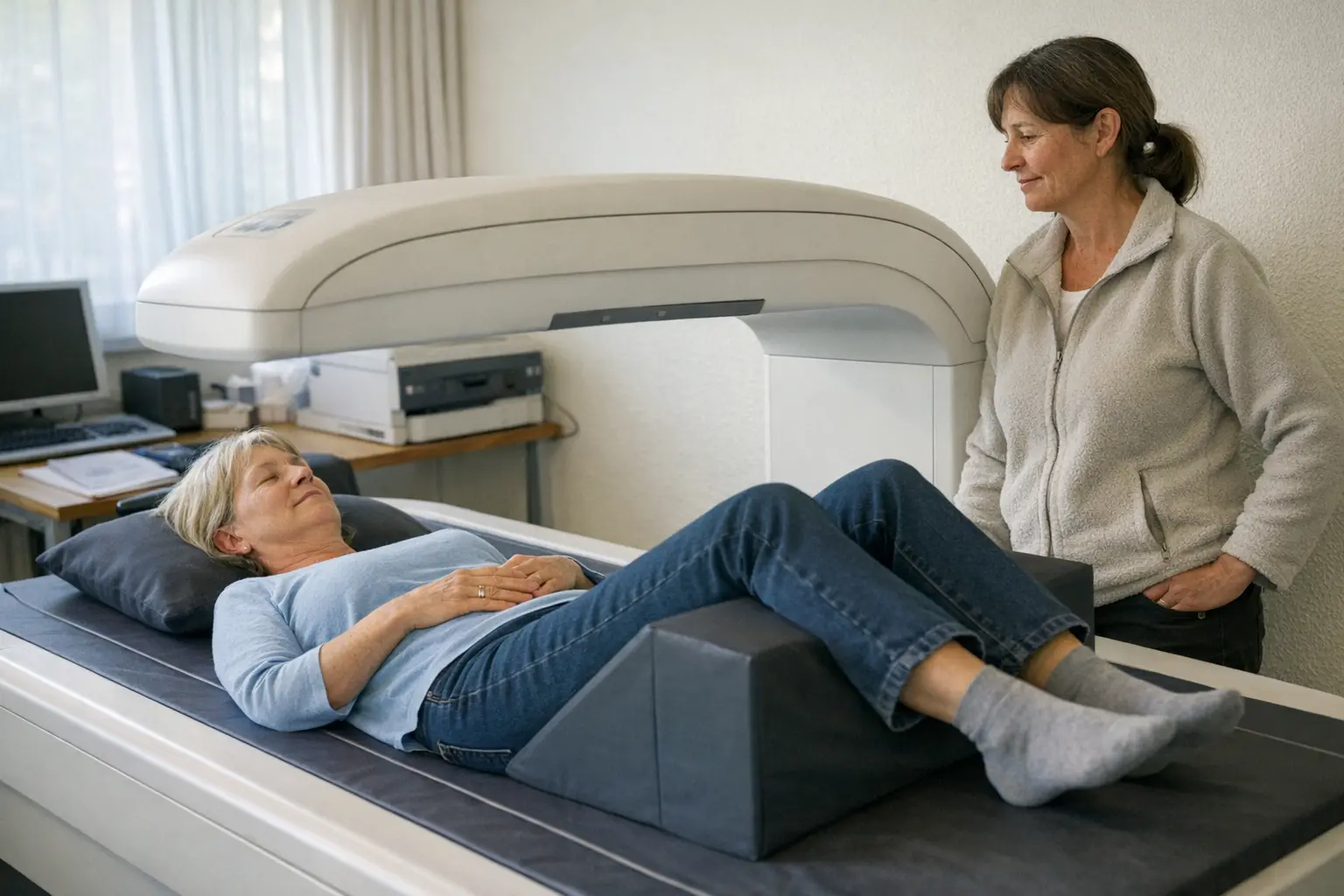
Bone density measurement (DXA scan) uses very low-dose X-rays to determine how strong your bones are. It is the standard examination for detecting osteoporosis — ideally before the first fracture occurs — and for monitoring the success of treatment.
Typical Areas of Use
- Investigation after a fracture without corresponding trauma
- Osteoporosis risk after menopause
- Monitoring during long-term corticosteroid treatment
- Investigation in case of loss of height or rounded back formation
- Follow-up monitoring during osteoporosis therapy
How It Works
- 1You lie comfortably and usually fully clothed on an examination table — no injection or tube is needed.
- 2A scanning arm moves slowly over the measurement regions, usually the lumbar spine and hip; you lie still for a few minutes.
- 3The device calculates your bone density from the measurements and compares it with reference values.
- 4Your doctor will discuss the result with you and evaluate it together with other risk factors — this determines whether and which treatment makes sense.
How to Prepare
- Wear comfortable clothing without metal parts such as belt buckles or zippers in the measurement area.
- Let us know if you have had an examination with contrast medium in the days beforehand — this can distort the measurement, so the appointment may need to be rescheduled.
- Calcium supplements should be omitted on the day of the examination depending on the practice's instructions — follow their directions.
Side Effects & Risks
- The radiation dose is extremely low — significantly lower than for an ordinary X-ray.
- The examination itself is painless and without noticeable side effects.
- The measured value alone does not constitute a diagnosis: only the medical evaluation together with age, medical history and other risk factors provides a complete picture.
There are no restrictions of any kind after the measurement. What matters most is the discussion of the results: if bone density is reduced, the doctor will discuss your options with you — ranging from exercise, strength training, and adequate calcium and vitamin D intake to medications that slow bone loss. Arrange an appointment promptly and do not wait if new, severe back pain occurs or you notice a marked loss in height — these can be signs of vertebral fractures. If numbness or weakness in the legs, or problems with urination, occur in addition, these symptoms need to be medically evaluated the same day.

Cost & Insurance
Swiss basic health insurance generally covers bone density measurement only when certain medical conditions are met — for example, after a fracture without adequate trauma, with known osteoporosis, or during long-term corticosteroid treatment. A purely preventive measurement without such risk factors often has to be paid for yourself. It is best to clarify the cost issue before the appointment with the practice or your health insurance provider; this information is provided for guidance only and without guarantee.
Frequently Asked Questions
- Does bone density measurement hurt?
- No, not at all. You simply lie still on a table for a few minutes while the scanning arm moves over you — you don't feel anything.
- How high is the radiation exposure?
- Very low — the dose from a DXA scan is far below that of an ordinary X-ray and in the range of natural radiation over just a few days. The examination can therefore be repeated without any problem.
- What does the T-score mean?
- The T-score compares your bone density with that of healthy young adults. Values below a certain threshold indicate reduced bone density or osteoporosis — your doctor will interpret what this means for you together with your other risk factors.
- Who should have a bone density measurement?
- Primarily people at increased risk: after a fracture without significant trauma, during long-term corticosteroid treatment, after early menopause, or with osteoporosis in the family. Your doctor will decide whether a measurement makes sense for you based on your risk profile.
- How often is the measurement repeated?
- Since bone density changes only slowly, follow-up examinations are typically meaningful only after around two years — depending on the findings and treatment, sometimes at longer intervals. Your doctor will determine the schedule individually.