
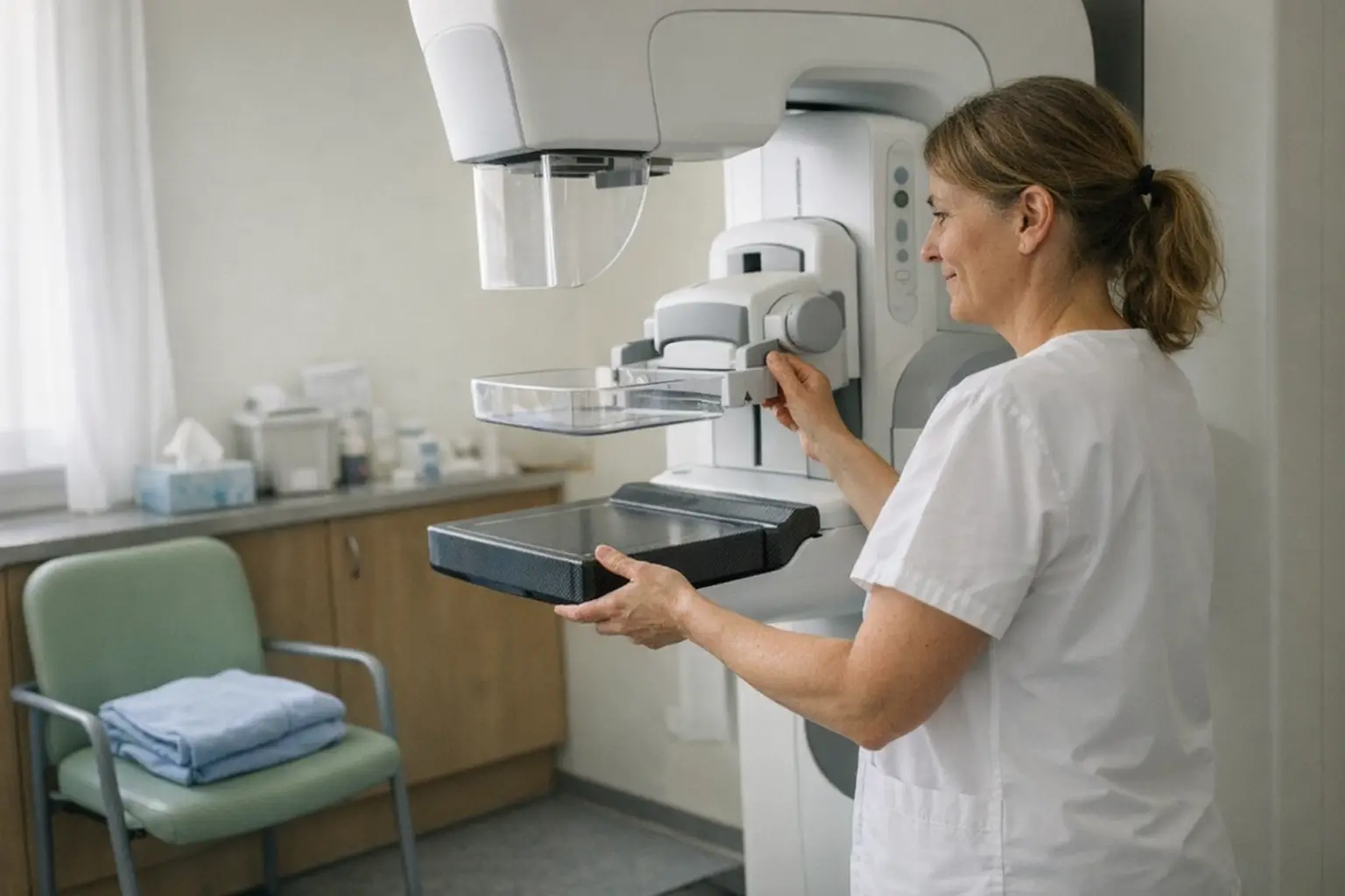
Mammography is an X-ray examination of the breast and the most important method for detecting breast cancer early — often long before a lump can be felt. It is used both for early detection from age 50 onwards and to investigate an abnormal finding on palpation.
Typical Areas of Use
- Breast cancer screening from age 50
- Investigation of a palpable lump in the breast
- Clarification of unclear findings from ultrasound
- Follow-up checks after breast cancer treatment
- More frequent monitoring in cases of increased familial risk
How It Works
- 1You undress your upper body; the technician positions one breast on the plate of the device.
- 2For the image, the breast is compressed between two plates for a few seconds — this is uncomfortable but necessary: the flatter the tissue, the more informative the image and the lower the radiation dose.
- 3As a rule, two images from different angles are taken per side; the whole process takes only a few minutes.
- 4The images are reported on by a radiologist — in screening programmes independently by two specialists. You receive the result in writing or in a consultation; if there are abnormalities, supplementary examinations such as ultrasound or a tissue sample follow.
How to Prepare
- Do not use deodorant, cream or powder on the breast and underarm area on the day of the examination — residues can mimic calcifications on the image.
- Two-piece clothing is practical, so that you only need to remove your top.
- Bring previous mammography images with you if these were taken elsewhere — comparison increases the diagnostic value.
- Before the menopause, the first half of the cycle is the most favourable time, as the breast is less sensitive then.
Side Effects & Risks
- The compression is uncomfortable to painful for a few seconds, but does not cause any damage.
- The radiation dose from modern equipment is low; according to current knowledge, the benefit of early detection clearly outweighs it.
- There are false-positive findings: abnormalities that turn out to be benign after further investigation — this can be worrying in the interim.
- Very dense breast tissue can make assessment more difficult; the doctor will often supplement the examination with an ultrasound in such cases.
- The screening also finds changes that would never have caused symptoms and are nevertheless treated (overdiagnosis). Benefits and harms can be compared in the information materials of the cantonal programme.
There are no restrictions after the mammogram; a slight feeling of tightness in the breast will resolve quickly. Wait calmly for the written report — being invited for further assessment does not yet mean a cancer diagnosis; in most cases, the finding turns out to be harmless. Regardless of your screening schedule, the following applies: arrange an appointment with your doctor promptly and do not wait if you feel a new lump, notice a change in the skin of the breast, a nipple that becomes retracted, or a nipple discharge.

Cost & Insurance
In cantons with an organised screening programme, women aged 50 and over are invited for a mammography every two years; the examination within the programme is exempt from the Franchise, though a co-payment may apply depending on the programme. Outside of programmes, mandatory health insurance covers the mammography if there is a medical justification, for example a palpable finding or an increased familial risk. If in doubt, please ask your health insurer or the cantonal programme; this information serves as guidance without guarantee.
Frequently Asked Questions
- Does a mammography hurt?
- The compression of the breast is uncomfortable for a few seconds, and for some women also painful — but it is over immediately afterwards. Before the menopause, it helps to schedule the appointment in the first half of the cycle, when the breast is less sensitive.
- Why does the breast have to be compressed so firmly?
- The flatter the tissue is spread out, the better the smallest changes can be detected — and the lower the necessary radiation dose. The compression is therefore not overzealousness, but a quality feature of the examination.
- From what age and how often is mammography advisable?
- In the cantonal programmes, women aged 50 and over are invited every two years. In cases of family history or symptoms, earlier or more frequent monitoring may be advisable — discuss your personal risk with your gynaecologist or GP.
- What does it mean if I am called in for further investigation?
- Initially, only that something needs to be looked at more closely — usually with ultrasound, additional images or a tissue sample. In the great majority of these investigations, no cancer is confirmed. The waiting period is stressful, but the thoroughness is worthwhile.
- What does mammography cost?
- In the cantonal screening programme, the Franchise is waived, so that at most you pay a small co-payment. Outside a programme, the Franchise and co-payment apply as usual, provided the examination is medically justified. Your health insurer can provide information in individual cases.