
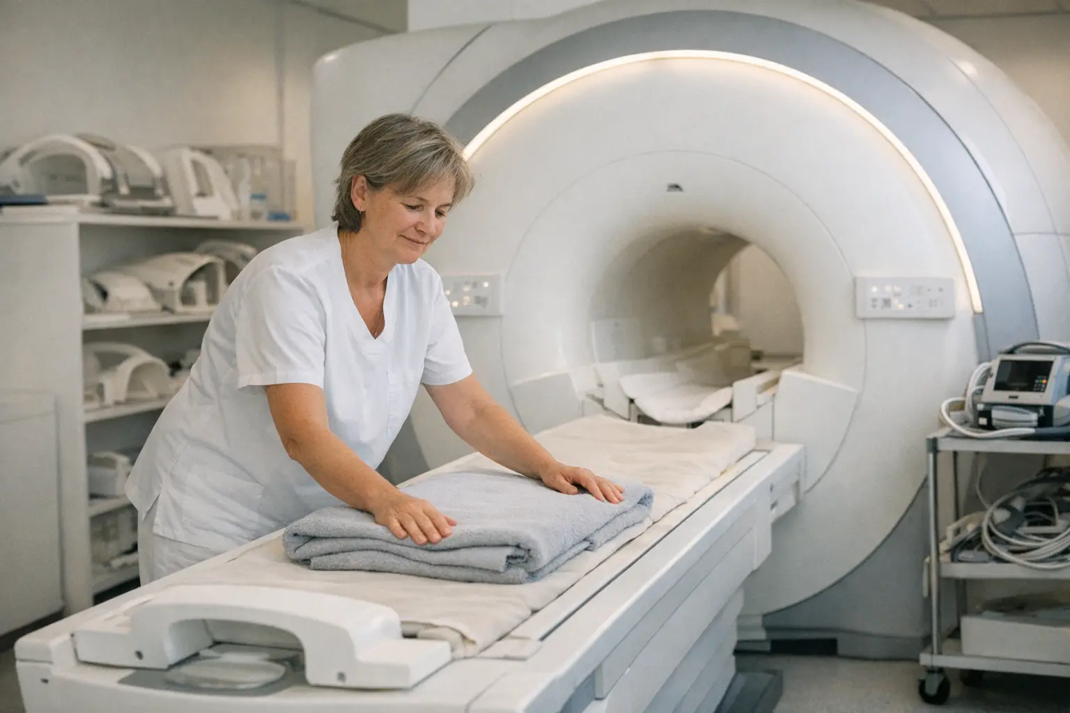
Magnetic resonance imaging (MRI) uses a strong magnetic field and radio waves to create detailed cross-sectional images of the inside of your body — entirely without X-rays. Soft tissues such as the brain, intervertebral discs, joints and internal organs are particularly well visualised. MRI helps your doctor get to the bottom of unclear symptoms with precision.
Typical Areas of Use
- Investigation of back and intervertebral disc problems
- Joint injuries such as meniscus or ligament tears
- Unclear headaches and neurological symptoms
- Assessment of internal organs
- Investigation of unclear findings from other examinations
How It Works
- 1Before the examination you complete a safety questionnaire: metal parts in the body, implants or a pacemaker must be reported without fail.
- 2You remove jewellery, watches, hearing aids and anything metallic; depending on the clinical question, access for contrast medium is placed in an arm vein.
- 3You lie on a table that moves into the tubular opening of the machine. During the imaging you will hear loud knocking sounds — you will be given hearing protection and an emergency call button with which you can alert the staff at any time.
- 4It is important to lie as still as possible so that the images are sharp. The team can see and hear you at all times.
- 5After the examination the images are reported by a radiologist; the result goes to your referring doctor, who will discuss it with you.
How to Prepare
- Leave jewellery and valuables at home if possible and wear clothing without metal parts.
- Bring implant cards with you (e.g. for pacemakers, artificial joints, stents) and report these when making your appointment.
- If you are prone to claustrophobia, mention it in advance — there are solutions, from music to open machines to a mild sedative. After a sedative you must not drive.
- Whether you need to come fasting depends on the examination; the institute's instructions are definitive.
Side Effects & Risks
- The examination itself is painless and does not involve X-rays.
- Some people find the narrowness of the tube and the loud noises unpleasant; hearing protection and support from the staff help with this.
- Contrast medium is usually well tolerated; allergic reactions occur rarely. Be sure to inform the radiology team about previous reactions to contrast medium, relevant kidney disease, and an existing or possible pregnancy.
- Certain implants or metal parts can limit or preclude the examination — that is why the safety questionnaire is so important.
After an MRI you can continue your everyday activities as normal. If you have received contrast medium, it helps to drink plenty of water throughout the day. After a sedative you must not drive for 24 hours and should arrange for someone to accompany you. Contact your doctor if you experience a rash, itching, swelling or breathing difficulties after contrast medium administration.

Cost & Insurance
An MRI referred by a doctor is generally covered by mandatory basic insurance; Franchise and deductible apply as usual. Without a doctor's referral — for example, as a purely voluntary check — you must expect costs of several hundred Swiss francs. If in doubt, ask your health insurance provider in advance; this information is a guide only and not guaranteed.
Frequently Asked Questions
- Does an MRI hurt?
- No, the examination is painless. The challenge is at most lying still and the loud noise — you will be given hearing protection for this. If contrast medium is injected, you will only feel the small needle stick in your arm vein.
- What should I do if I have claustrophobia?
- Definitely mention it when making your appointment. Many institutes offer more spacious machines, music or support from staff; in severe cases a mild sedative can help. However, you must not drive afterwards.
- How long does an MRI take?
- Depending on the region being examined, approximately 20 to 60 minutes. Allow a good hour in total including preparation and changing.
- Can I have an MRI with an implant?
- Many modern implants are MRI-compatible, but this must be checked in advance. Bring your implant card with you and report pacemakers, cochlear implants, metal parts or fragments when registering.
- Can I drive after the MRI?
- Yes — unless you have received a sedative. In that case you must not drive any vehicle for 24 hours and should arrange to be collected.
- What is the difference between MRI and CT?
- MRI uses magnetic fields and shows soft tissues such as intervertebral discs, ligaments or the brain particularly well. CT uses X-rays, is faster and is particularly suitable for bones, lungs and emergency situations. Your doctor will choose the method that suits your clinical question.