
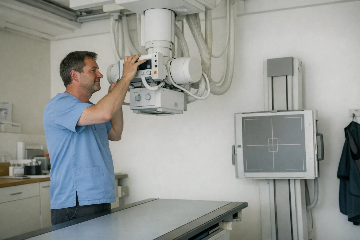
X-ray imaging is the classic and fastest form of imaging: within a few minutes, an image of bones, joints or the lungs is created. It is often the first step when investigating a broken bone, joint wear or pneumonia.
Typical Areas of Use
- Suspected bone fracture after a fall or accident
- Investigation of joint wear (osteoarthritis)
- Lung examination, for example in cases of suspected pneumonia
- Monitoring the healing process after a fracture
- Assessment of misalignments
How It Works
- 1You remove jewellery and metal objects in the area being examined; depending on the region, you may need to remove the relevant item of clothing.
- 2The specialist positions you standing, sitting or lying down and directs the device precisely at the region to be examined.
- 3For the exposure, you hold still for a few seconds — for lung images, you briefly hold your breath. Usually two images are taken from different angles.
- 4The images are immediately available digitally and are assessed by the doctor and discussed with you.
How to Prepare
- No special preparation is necessary; practical clothing that can easily be removed in the examination area is helpful.
- You must report a possible pregnancy before the examination.
Side Effects & Risks
- The radiation dose from a single exposure is low — for many exposures, roughly equivalent to what you would absorb from a few days of natural background radiation.
- During pregnancy, X-rays are only taken if absolutely necessary; therefore always report a possible pregnancy.
- Soft tissues such as ligaments, tendons or intervertebral discs are barely assessable on X-ray images — sometimes ultrasound, CT or MRI may be needed as a supplement.
After the X-ray, there is nothing special to watch out for — you can return to your daily routine immediately and are fully fit to drive. The only important thing is to discuss the findings: clarify with your doctor what the image shows and what steps may follow. Contact your doctor if your symptoms persist or worsen despite an unremarkable image.

Cost & Insurance
X-rays ordered by a doctor are usually covered by basic health insurance; the deductible and co-payment apply as usual. Many GP and orthopaedic practices can perform X-rays directly on site. If in doubt, ask your health insurance provider; this information is for guidance only and without guarantee.
Frequently Asked Questions
- Does an X-ray hurt?
- No, you don't feel anything at all from the exposure itself. Only the positioning may be uncomfortable if the region being examined is already painful — tell the specialist, they will help you with positioning.
- How harmful is the radiation?
- The dose from a single exposure is low and comparable to a few days of natural background radiation. Nevertheless, X-rays are only taken if the image answers a specific question — and during pregnancy only if unavoidable.
- How quickly will I have the result?
- The images are available digitally immediately. In many practices, the doctor discusses the findings with you directly afterwards.
- Can you see ligaments and tendons on an X-ray image?
- Barely — X-rays primarily show bones. For ligaments, tendons, cartilage or intervertebral discs, ultrasound or MRI are more suitable. Your doctor will decide which imaging method fits your case.
- Do I have to go to hospital for an X-ray?
- Not necessarily. Many GP practices, orthopaedic practices and X-ray institutes perform examinations directly on site, often without long waiting times.